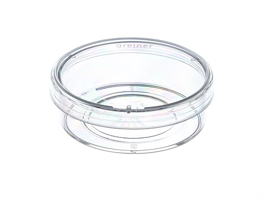
CELLview™, boite culture cellulaire, PS,
35x10mm, Fd en verre, 1 compartiment, stérile, 10 PCS./sachet

Référence produit: 627861
Informations principales
| Description du produit: |
CELLview™, boite culture cellulaire, PS, 35x10mm, Fd en verre, 1 compartiment, stérile, 10 PCS./sachet |
|---|---|
| Stérile: | sterile |
| Conditionnement: | 40 |
| Sous conditionnement : | 10 |
Emballage
| Poids emballage: | 0,27 kg |
|---|---|
| Dimension emballage: | 195 x 145 x 125 mm |
| Conditionnement: | 40 |
| Sous conditionnement : | 10 |
| PAL: | 22800 |
Informations détaillées
| Volume de remplissage (ml)\n: | 0.00 |
|---|
CELLview - Boîte de Culture Cellulaire à Fond Verre
- Analysé pour detection ADNase, ARNase et ADN humain
- Exempt de substances cytotoxiques
- Caractéristiques du fond verre:
- Verre borosilicate achromatique à haute transparence, hydrolytique de classe I (DIN ISO 719)
- Epaisseur du verre 175 µm +/- 15 µm
- Transmission spectrale maximale, sans auto fluorescence
- Avantages:
- La version compartimentée permet l’analyse en multiplex
- Fond verre intgré pour und planéité maximale
- Nombre de compartiments: 1
- Diamètre: 35 mm; hauteur: 10 mm
- Surface de culture: 8,7 cm²
- Volume total: 10 ml
- Volume de travail: 5 ml
- Stérile
Videos
Drug treatment during live cell imaging
A multi-position time-lapse experiment was started and after acquiring six time points every two minutes drugs were added to the different wells as indicated:
- Video 1 - control (no drugs added)
In steady-state the Golgi apparatus is relatively stable on light microscopy level. The shape changes only slowly during the time of the experiment when observing control cells. Also the number of Golgi fragments visible by light microscopy resolution is relatively constant over time.
- Video 2 - Nocodazole added, final concentration 10 µM
Nocodazole treatment induces, fragmentation of the Golgi apparatus. The onset of fragmentation starts 10 to 15 minutes after addition of the drug. The onset of fragmentation differs between individual cells. Fragmentation of the central Golgi to many distributed ministacks is the final phenotype of microtubule depolymerization after three hours.
- Video 3 - Latrunculin B added, final concentration 1 μM
Actin depolymerization by Latrunculin B influences the shape of the Golgi from relatively thin elongated to a rounded up and compact appearance. After 10 to 20 minutes differences in the Golgi morphology became first visible and after approximately one hour the Golgi rearrangement was completed.
- Video 4 - Brefeldin A added, final concentration 5 μg/ml
Block of export from the endoplasmatic reticulum (ER) by Brefeldin A leads to a rapid redistribution of the Golgi compartment to the ER by retrograde transport. This effect is often completed within 5 minutes.
Performing these experiments in parallel in CELLview dishes with four compartments it is possible to directly compare the speed and timing of drug effects on the Golgi apparatus. Brefeldin A affects Golgi morphology much faster than Nocodazole and Latrunculin B, which both induces first changes in the range of 10-20 minutes.
Documents associés
Application Note: siRNA dependent gene silencing in HeLa cells cultivated on
Notes d'application / PDF, 508 KB
Notes d'application / PDF, 508 KB
Forum No 12: Advanced TC™: An innovative surface improving cellular assays
Publications scientifiques / PDF, 607 KB
Publications scientifiques / PDF, 607 KB
Application Note: Cultivation and Differentiation of Human Adipose Derived Mesenchymal Stem Cells with CELLSTAR® and CELLCOAT® Cell Culture Products
Notes d'application / PDF, 539 KB
Notes d'application / PDF, 539 KB
Application Note: Improved Cultivation and Differentiation of Embryonic Stem Cells
Publications scientifiques / PDF, 2 MB
Publications scientifiques / PDF, 2 MB
C'est le moyen le plus simple de tout savoir. Nous serions ravis de vous compter parmi notre communauté d'abonnés.