


THINCERT CELL CULTURE INSERT FOR 6 WELL PLATES, TC, STERILE, TRANSPARENT MEMBRANE (PET), PORE DIAMETER: 3 µm, 4 MULTIWELL PLATES/BOX, 24 THINCERT INSERTS/BOX
For advanced cell and tissue culture applications, Greiner Bio-One offers an extensive family of membrane supports - ThinCert®. They are available in 6, 12 and 24 well sizes and can be combined with six different membrane types of different pore sizes and density. They are compatible with CELLSTAR® standard cell culture multiwell plates.
ThinCert®Plates have been optimized for use with ThinCert® cell culture inserts. The deep wells of this plate allow the application of a larger medium volume during air-lift culture. As a consequence, fewer medium changes are required and the amount of manual work is significantly reduced. These are available in 6 well and 12 well formats.
The ThinCert® cell culture inserts are suitable for a wide range of applications including:
Material:
ThinCert® cell culture inserts are produced from high-grade clear polystyrene housings, and polyethylene terephthalate (PET) capillary pore membranes. Both materials, polystyrene and PET, are USP class VI certified and cell culture compatible. The coupling between the housing and the membrane is achieved using an automated process. This produces an extremely strong and robust seal without compromising or weakening the membrane in any way.
The membranes undergo a physical surface treatment to optimise cellular adherence and growth characteristics. All the capillary pores in a membrane exhibit a high degree of uniformity in diameter. This uniformity ensures reliable and consistent exchange rates between the two compartments and thus provides reproducibility when conducting multiple experiments.
Kits:
The cell culture inserts are pre-packed together with the requisite number of plates. The automated production process includes double optical control of each insert produced, ensuring that any biological contamination is avoided. The sterility of the single blisterpacked inserts and multiwell plates is ensured by irradiation.
For light or electron microscopic examinations, the membranes can be easily detached from the housing using a scalpel. Even when detached, the membrane remains flat without unrolling, greatly simplifying further steps. Due to its high chemical resistance to various solvents, the membrane is suitable for numerous cell fixation protocols. Due to their special suspension, ThinCert® cell culture inserts keep a distance to the well bottom at all times, which means that cells cultured there are always protected from damage. A safe minimum distance to the side walls is always maintained with the help of distance holders, which prevents capillary suction between the inner wall of the well and the outer wall of the insert. This means that mass transfer between the well and ThinCert® takes place exclusively via the porous membrane. ThinCert® cell culture inserts sit eccentrically in the wells and deflect upwards when a pipette is inserted. When the pipette is removed again, the ThinCert® automatically resume their original position. This feature is called self-lift geometry.
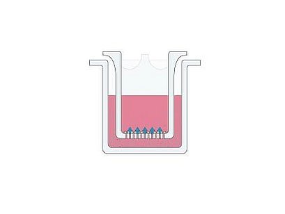
Migration/ Invasion assay
Cell migration plays a significant role in physiological and pathological processes during embryonic development. As well as wound healing, immune response, inflammation, and tumorigenesis. The filter assay is a standard in vitromodel used to study cell migration. It is performed in cell culture inserts with 8.0 μm pores of the ThinCert® membrane. Cell migration is supported and involved from the upper compartment towards a chemo-attractant source in the lower compartment.
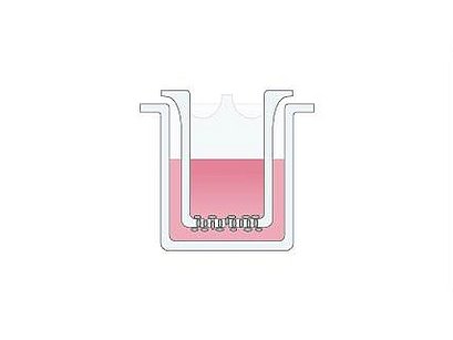
Co-Culture
Co-culture involves the study of immune cell interactions. Additionally, it includes other diverse applications such as the stimulation of cell proliferation, the maintenance of cell differentiation, and the restoration of heterocellular functions in vitro (e.g. bloodbrain-barrier). With ThinCert® cell culture inserts, cells may be seeded in the upper and lower compartment. The 0.4 µm or 1.0 µm pores of the ThinCert® membrane allow the exchange of molecules between the two cell populations.
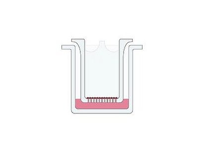
Transport Studies
Transport studies are among the most frequent applications of cell culture inserts. The goal is to reconstruct a functional epithelium from individual cells. Additionally, an active transport of substances from one compartment through the epithelium into the other compartment will be investigated. ThinCert® cell culture inserts with 0.4 µm pores and translucent membranes are recommended for transport assays.
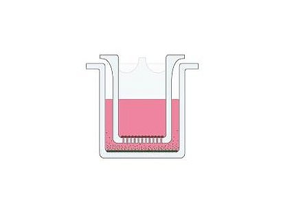
Organotypic and air-lift-culture
In organotypic culture a tissue can be kept alive for prolonged periods. While in tissue reconstruction the tissue is de novo generated from single cells. Both procedures use cell culture inserts with 0.4 to 3.0 µm pores. The tissue growth in vitro at the air-liquid-interface without limitations from gas exchange. Furthermore, for some tissue types, the direct exposure of the cultivated cells to the surrounding atmosphere serves as an indispensable differentiation stimulus.
Cell biology research and cell-based screening are going through an exciting period of development; he broad adoption of 3D cell culture. We at Greiner Bio-One are very enthusiastic about it and want to share with you methods and techniques that are producing results.
THINCERT CELL CULTURE INSERT FOR 6 WELL PLATES, TC, STERILE, TRANSPARENT MEMBRANE (PET), PORE DIAMETER: 3 µm, 4 MULTIWELL PLATES/BOX, 24 THINCERT INSERTS/BOX

THINCERT CELL CULTURE INSERT FOR 24 WELL PLATES, TC, STERILE, TRANSPARENT MEMBRANE (PET), PORE DIAMETER: 3 µm, 2 MULTIWELL PLATES/BOX, 48 THINCERT INSERTS/BOX


THINCERT CELL CULTURE INSERT FOR 6 WELL PLATES, TC, STERILE, TRANSPARENT MEMBRANE (PET), PORE DIAMETER: 1 µm, 4 MULTIWELL PLATES/BOX, 24 THINCERT INSERTS/BOX

THINCERT CELL CULTURE INSERT FOR 6 WELL PLATES, TC, STERILE, TRANSLUCENT MEMBRANE (PET), PORE DIAMETER: 3 µm, 4 MULTIWELL PLATES/BOX, 24 THINCERT INSERTS/BOX

THINCERT CELL CULTURE INSERT FOR 6 WELL PLATES, TC, STERILE, TRANSLUCENT MEMBRANE (PET), PORE DIAMETER: 8 µm, 4 MULTIWELL PLATES/BOX, 24 THINCERT INSERTS/BOX

THINCERT CELL CULTURE INSERT FOR 6 WELL PLATES, TC, STERILE, TRANSLUCENT MEMBRANE (PET), PORE DIAMETER: 0,4 µm, 4 MULTIWELL PLATES/BOX, 24 THINCERT INSERTS/BOX

THINCERT CELL CULTURE INSERT FOR 6 WELL PLATES, TC, STERILE, TRANSPARENT MEMBRANE (PET), PORE DIAMETER: 0,4 µm, 4 MULTIWELL PLATES/BOX, 24 THINCERT INSERTS/BOX

THINCERT CELL CULTURE INSERT FOR 24 WELL PLATES, TC, STERILE, TRANSPARENT MEMBRANE (PET), PORE DIAMETER: 1 µm, 2 MULTIWELL PLATES/BOX, 48 THINCERT INSERTS/BOX

THINCERT CELL CULTURE INSERT FOR 24 WELL PLATES, TC, STERILE, TRANSLUCENT MEMBRANE (PET), PORE DIAMETER: 3 µm, 2 MULTIWELL PLATES/BOX, 48 THINCERT INSERTS/BOX

THINCERT CELL CULTURE INSERT FOR 24 WELL PLATES, TC, STERILE, TRANSLUCENT MEMBRANE (PET), PORE DIAMETER: 8 µm, 2 MULTIWELL PLATES/BOX, 48 THINCERT INSERTS/BOX

THINCERT CELL CULTURE INSERT FOR 24 WELL PLATES, TC, STERILE, TRANSLUCENT MEMBRANE (PET), PORE DIAMETER: 0,4 µm, 2 MULTIWELL PLATES/BOX, 48 THINCERT INSERTS/BOX

THINCERT CELL CULTURE INSERT FOR 24 WELL PLATES, TC, STERILE, TRANSPARENT MEMBRANE (PET), PORE DIAMETER: 0,4 µm, 2 MULTIWELL PLATES/BOX, 48 THINCERT INSERTS/BOX


THINCERT CELL CULTURE INSERT FOR 12 WELL PLATES, TC, STERILE, TRANSPARENT MEMBRANE (PET), PORE DIAMETER: 1 µm, 4 MULTIWELL PLATES/BOX, 48 THINCERT INSERTS/BOX

THINCERT CELL CULTURE INSERT FOR 12 WELL PLATES, TC, STERILE, TRANSPARENT MEMBRANE (PET), PORE DIAMETER: 3 µm, 4 MULTIWELL PLATES/BOX, 48 THINCERT INSERTS/BOX

THINCERT CELL CULTURE INSERT FOR 12 WELL PLATES, TC, STERILE, TRANSLUCENT MEMBRANE (PET), PORE DIAMETER: 3 µm, 4 MULTIWELL PLATES/BOX, 48 THINCERT INSERTS/BOX

THINCERT CELL CULTURE INSERT FOR 12 WELL PLATES, TC, STERILE, TRANSLUCENT MEMBRANE (PET), PORE DIAMETER: 8 µm, 4 MULTIWELL PLATES/BOX, 48 THINCERT INSERTS/BOX

THINCERT CELL CULTURE INSERT FOR 12 WELL PLATES, TC, STERILE, TRANSLUCENT MEMBRANE (PET), PORE DIAMETER: 0,4 µm, 4 MULTIWELL PLATES/BOX, 48 THINCERT INSERTS/BOX

THINCERT CELL CULTURE INSERT FOR 12 WELL PLATES, TC, STERILE, TRANSPARENT MEMBRANE (PET), PORE DIAMETER: 0,4 µm, 4 MULTIWELL PLATES/BOX, 48 THINCERT INSERTS/BOX